Infrastructure
BIOLOGICAL PHYSICS LAB
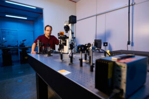
OPTICAL TWEEZERS
Description: experimental technique that allows the manipulation of systems and / or objects with typical size in the range of nano to micrometers. Uses a laser strongly focused by an objective lens to exert force on objects. The technique allows one to manipulate, move, stretch, compress and rotate these objects by exerting forces in the typical range of femtoNewtons to hundreds of picoNewtons.
Available equipment: 03 inverted optical microscopes (Ti-S, Ti-U and Ti-S2, Nikon); 01 ytterbium-doped fiber IR laser (1064 nm, 5 W) ; 02 solid state IR lasers (1064 nm, 2 W); 01 multi-wavelength laser; 02 spatial light modulators; 02 nanometric PZT drive systems; 02 optical tables; optical and mechanical elements: lenses, mirrors, filters, positioning systems, etc.; image capture systems with various CCD and CMOS cameras.
MAGNETIC TWEEZERS
Description: experimental technique that allows the manipulation of systems and / or objects with a typical size in the nano / micrometer scale, with the ease of applying torques and exerting constant force on these systems. It uses a magnetic field generated in the sample region (which is usually mounted under an optical microscope) to exert force on paramagnetic particles.
Equipment available: magnets, coils and current sources of various characteristics. It shares the same microscopes and opto-mechanical elements of the optical tweezers.
CELL CULTURE ROOM
Description: complete infrastructure for growing normal and cancer cell cultures, which are used in our experiments for studying the dynamics of migration, aggregation and membrane activity of these cells.
Available equipment: clean room for preparation of cell cultures; 01 biological microscope Nikon BioStation with temperature, humidity and CO2 control for experiments with living cells, 01 CO2 incubator, 01 laminar flow, 01 microcentrifuge, 02 cryogenic canisters for cell storage, 02 inverted microscopes for videomicroscopy (Ti-S and TS-100, Nikon); 01 inverted microscope for routine use.
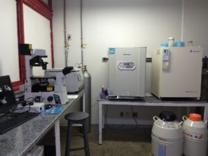
Cell culture room.
GEL ELECTROPHORESIS ROOM
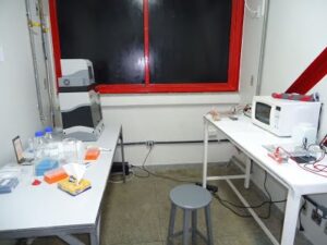
Description: experimental technique that allows one to study the mobility of DNA molecules as well as DNA-ligand complexes in agarose or polyacrylamide gels by applying an electric field. It can be used to separate DNA fragments by size and to infer structural changes on the molecules.
Available equipment: 03 chambers (02 horizontal and 01 vertical), 03 power sources for electrophoresis; 01 photodocumentation system.
FLUORESCENCE MICROSCOPY
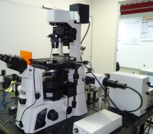
Description: experimental technique that allows one to view and follow processes in living cells and molecules (DNA, proteins, etc.) properly labeled with a fluorescent dye.
Available equipment: 01 fluorescence microscope Nikon BioStation, 01 inverted optical microscope Nikon Ti-U with EPI-fluorescence.
OTHER EQUIPMENT AVAILABLE IN ASSOCIATED LABS
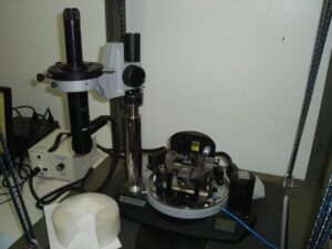
ATOMIC FORCE MICROSCOPE
Description: allows one to view systems with sizes in the nanometer scale (properly deposited on a substrate), by using a scanning probe.
DYNAMIC LIGHT SCATTERING (DLS)
Description: allows one to infer the typical sizes and associated fluctuations for small particles (DNA, proteins…), by analyzing of the light backscattered by these systems.

